Postdoctoral Scholar Spotlight: Marina Ayad
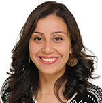 Marina Ayad, PhD
Postdoctoral Scholar
Department of Pathology
What projects are you currently working on or interested in?
I am currently working on developing AI tools to study the brain tumor microenvironment, with a focus on meningioma and glioblastoma. Although both tumors originate in the brain, they have highly different pathologies and clinical outcomes. Meningiomas are mostly considered benign tumors, however, predicting their recurrence based on histopathology alone remains challenging. My work on meningioma focused on developing interpretable AI models that not only predict recurrence risk but also identifies tissue patterns associated with higher risk tumors.
One interesting outcome from this project was that AI-assisted quantification of the tissue features achieved higher predictive accuracy than conventional WHO grading criteria, highlighting the potential of AI to both enhance current diagnostic criteria and uncover clinically relevant patterns that may otherwise go unrecognized.
I am also studying the tumor microenvironment in glioblastoma which is a highly aggressive tumor that inevitably recurs after treatment. I am specifically focusing on the extracellular matrix, the network of proteins that surrounds cells, and how it influences therapy resistant cancer stem cells that may drive tumor recurrence. For this study, I am analyzing spatial transcriptomics datasets to characterize tumor microenvironment interactions at a spatial resolution. My long-term goal is to translate these insights into AI tools that assist pathologists detect and interpret subtle tissue patterns, such as extracellular matrix remodeling, from routine acquired pathology slides.
What is a cause that you are passionate about?
I am intrigued by the idea that a pathology slide contains a record of a tumor’s history and evolution. The tissue patterns that pathologists observe under the microscope are the result of multiple layers of biological and genetic changes that unfolded over time. While AI in computational pathology has already shown significant value in clinical diagnostics, many models still function as black boxes. They often provide limited insight into tissue patterns and underlying biological processes that drive their predictions.
I am interested in developing AI tools that can help decode these patterns, revealing insights into how tumors evolve, recur and respond to therapy. By connecting tissue patterns to biological insights, these models would enable pathologists and clinicians to use information embedded within routine tissue samples not only for diagnosis, but also to make more informed treatment decision.
What advice would you give to a student wanting to get into this field of study?
I think there are two key aspects to keep in mind: mentorship and mastering the fundamentals.
Mentorship is a key aspect of a becoming a researcher. There is a huge value in being mentored by someone who is not just a great scientist but also has wisdom to navigate questions that have never been answered before. A great mentor provides strategic insight, helping you recognize when to keep pursuing a challenging problem and when it’s time to change direction.
The second piece of advice is to focus on understanding the core concepts before jumping into large-scale projects. In the era of AI, building models has become easier than it was a couple of years ago. However, meaningful scientific progress requires more than applying the latest tools. It requires understanding strong foundations in both biology and computational methods to know when a method is appropriate, recognize its limitations and identify the questions that are worth pursuing.
What are your plans for the future?
I will be joining Moffitt Cancer Center as an assistant professor in the Department of Translational Pathology. My lab will focus on studying the tumor tissue architecture and how it reflects underlying biological processes that drive disease progression and clinical outcomes. To achieve this goal, we will develop interpretable AI models that can generate novel biological hypotheses, integrate histology with spatial molecular imaging technologies, and model the tumor microenvironment, including subtle patterns such as the extracellular matrix organization. Ultimately, my vision is to leverage patient-derived tissue resources to develop more precise diagnostic, prognostic and therapeutic strategies for cancer patients.