Preserving the X-rays That Helped Transform Healthcare
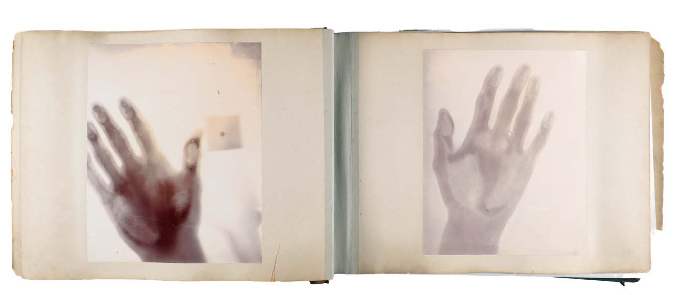
As an orthopaedic surgeon, Joseph J. Gugenheim, ’72 MD, spent his career relying on medical imaging to care for patients. So when he learned that Galter Health Sciences Library at Northwestern University Feinberg School of Medicine kept an album documenting some of the earliest physician-produced X-rays in the U.S., the collection resonated with both his professional experience and lifelong interest in medical history.
Created in 1896, just weeks after X-rays were introduced to the world, the images capture the dawn of a technology that would become indispensable to modern patient care. “Northwestern has always been an innovator,” Dr. Gugenheim said, explaining that the collection encapsulates that spirit of discovery so ingrained at Feinberg.
Recognizing the collection’s historical significance, Dr. Gugenheim made a generous gift to support its conservation and digitization. The album contains 18 X-ray images and is believed to include the first X-rays made in Chicago as well as some of the earliest physician-produced X-rays in the country, according to Katie Lattal, head of Special Collections at Galter Library. “These X-rays are really special—to Chicago, to radiology, and to medicine,” Lattal said.
The album documents the work of James Burry, 1875 MD, a Chicago surgeon and alumnus of Chicago Medical College, which later evolved into Northwestern’s medical school. Dr. Burry and a team of engineers at Western Electric Co. used the new technology to photograph hands and study exposure times, creating images that today might appear crude but were groundbreaking at the time. The collection also includes evidence of what is believed to be one of the first surgeries guided by prior X-ray imaging, held at Mercy Hospital (now Insight Hospital and Medical Center) and reported in local and national media.
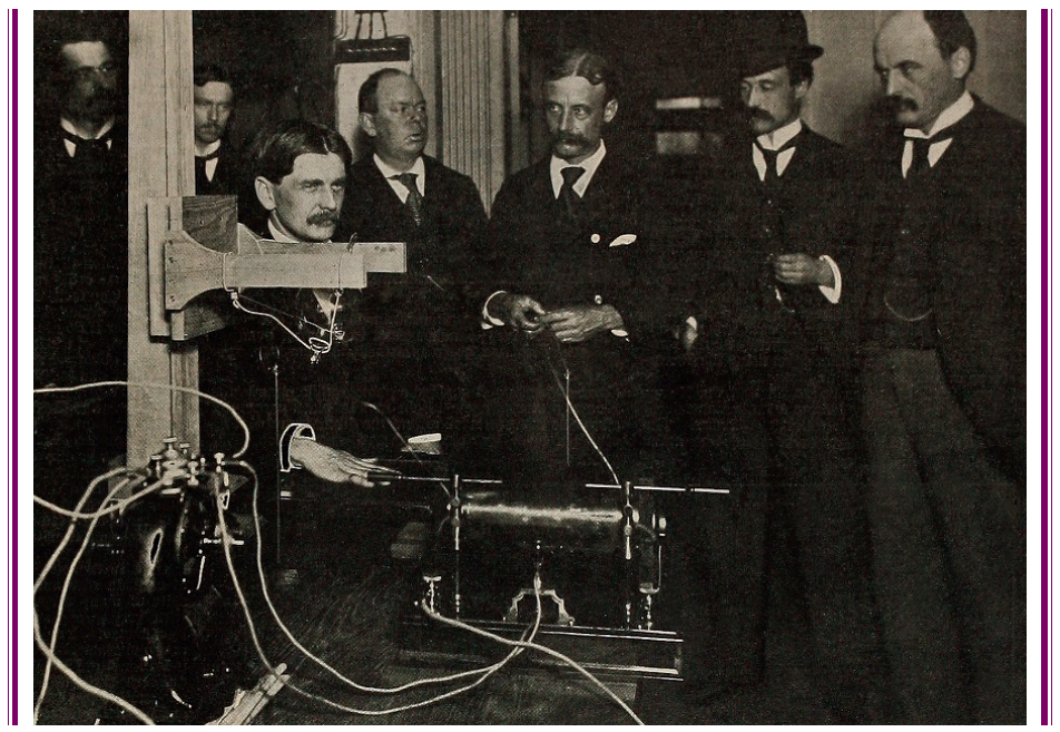
In 1896, producing an X-ray was a far more laborious process than the near-instant imaging physicians use today. Early X-rays were recorded on photographic plates and later developed much like photographs in a darkroom. The process required chemicals, silver-based film, and careful handling. The album at Galter Library evinces that era of experimentation, when physicians and engineers were still learning how to create clear images and standardize the technology. The X-rays featured in the collection required 30-45 minutes of exposure.
By contrast, Dr. Gugenheim said, modern hospitals have largely replaced film with digital imaging systems. During much of his career, healthcare institutions maintained vast X-ray film storage rooms containing files that sometimes had to be physically transported when patients sought care elsewhere. The films could be misplaced, damaged, or lost, creating challenges for both patient care and recordkeeping. Today, digital images can be captured, stored, and transmitted electronically within seconds, allowing physicians across departments—or even across healthcare systems—to access the same information almost immediately.
More than 130 years later, the fragile photographic prints in Galter Library’s collection have faded and deteriorated. Dr. Gugenheim’s gift will allow conservators to stabilize the album, create high-resolution digital copies, and produce facsimiles for study, protecting the originals while expanding access for scientists.
By preserving these pioneering images, Dr. Gugenheim said he hopes his gift will ensure that future generations can explore one of the earliest moments in the evolution of medical imaging.
“I feel like I and others have an obligation to continue showing the high quality of our medical school. It’s been a leader since 1859, and this collection further documents that,” he said.
For more information about supporting Galter Library, please contact David McCreery at david.mccreery@northwestern.edu or 312-503-6099.
This story was published in the Fall 2026 issue of The Philanthropist, a newsletter for supporters and friends of Northwestern University Feinberg School of Medicine. Read past issues here.