Investigator Resources
The Stephen M. Stahl Center for Psychiatric Neuroscience is home to a wide range of expertise related to the study of mental health and brain function. Contact us for additional support or requests.
Approaches
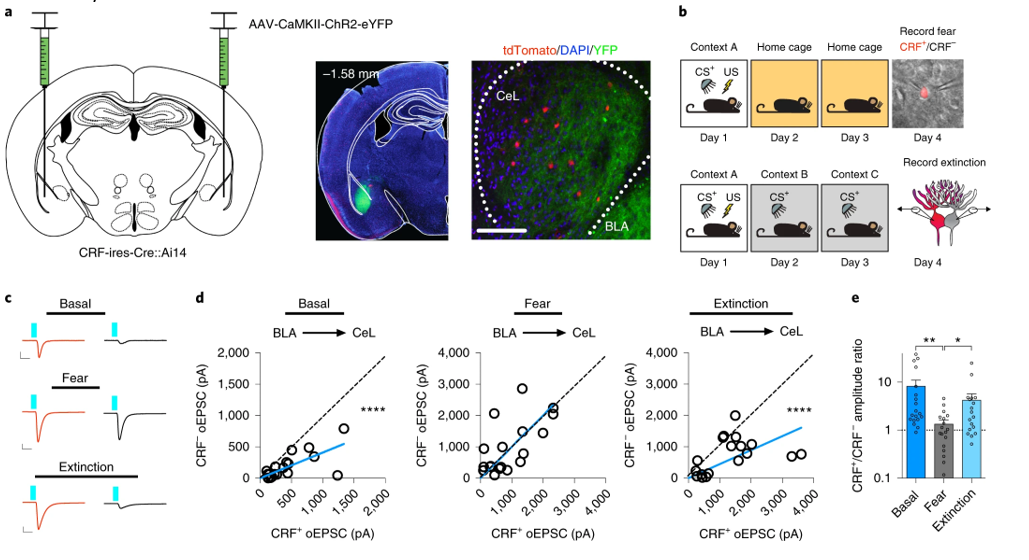
Brain Slice Electrophysiology
Center investigators have considerable expertise in electrophysiological approaches to studying neuronal circuit function. Combining whole-cell patch-clamp methods with optogenetics and genetically-driven reporter models allows for circuit- and cell-type specific interrogation at the synaptic level. Understanding pharmacologically and experience-dependent adaptations in synaptic and circuit function related to disease phenotypes could provide novel insights into circuit-level pathophysiological mechanisms of psychiatric disorders and drug efficacy.
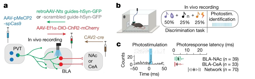
In Vivo Electrophysiology
The ability to record large-scale electrical signals in distinct brain regions and coupling between brain regions represents a major advance in neuroscience. Center investigators utilize advances in vivo neuronal activity measures, including photo tagging and cell-type specific analyses combined with cutting-edge analytical and statistical approaches, to elucidate how distinct cell types represent and encode cognitive, emotional and valence-related processes in the brain.
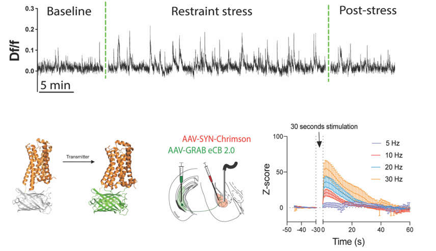
Fiber Photometry
Fiberoptic-based analysis of florescent signaling in awake, behaving mice has recently enabled major advances in understanding how specific circuits and cell types regulate distinct behavioral processes. In addition, exponential growth of fluorescent biosensors has expanded our ability to monitor release of neurotransmitters within distinct neural circuits with high temporal precision for the first time. Center investigators are on the leading edge of fiber-based methods development and application to high-impact translational calcium imaging (neurons utilize intracellular calcium dynamics for a variety of purposes including coincidence detection, membrane-to-nucleus signaling and synaptic plasticity). New advances in calcium imaging in awake, freely moving animals has allowed for an unprecedented level of analysis from identified cell types and neural pathways. Center investigators are highly knowledgeable about in vivo calcium imaging approaches and the application of this technology to answer important questions in translational psychiatry.
Behavioral Phenotyping
Analysis of behavioral phenotypes is a critical aspect of translational neuroscience research, and our investigators have extensive experience in mouse behavioral phenotyping. The center houses equipment for a variety of behavioral analyses compatible with in vivo brain activity monitoring, ranging from classical and operant conditioning to social behavior and pain assays
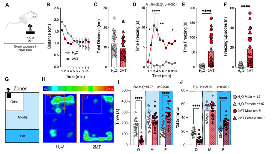
Pictured: Example of mouse behavioral phenotyping in response to predator odor exposure. Graphs depict increases in fear behavior and avoidance of predator odor but not water presentation.
Optogenetics to Manipulate Neuronal Activity
Use of light-activated ion channels to interrogate neural circuit function has revolutionized systems and translational neuroscience. Center investigators utilize in vivo and ex vivo optogenetic approaches to understand how neural circuits control behavioral phenotypes relevant to psychiatric disease in order to ultimately gain insight into neural circuit-level pathophysiological mechanisms underlying psychiatric illness.
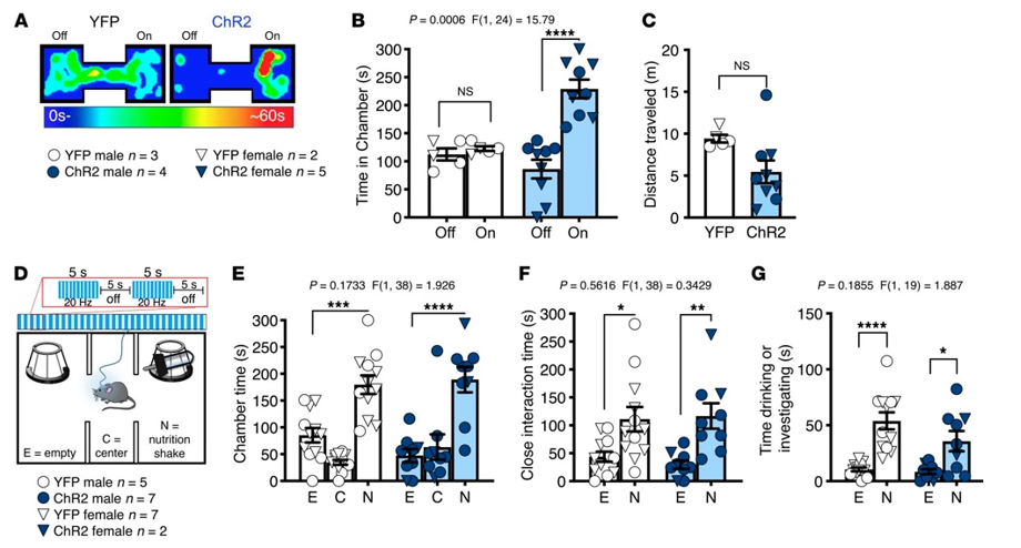
Pictured: Excitatory optogenetic activation of amygdala inputs to the nucleus accumbens produced a real-time place preference but did not affect preference for natural reward in a three-chamber test.
Neurochemistry
Analysis of neurotransmitter release via use of microdialysis, for example, represents a cornerstone approach for translational psychiatry and psychopharmacology research. The center houses state-of-the-art mass spectrometry-based neurochemistry resources for our investigators and collaborators.
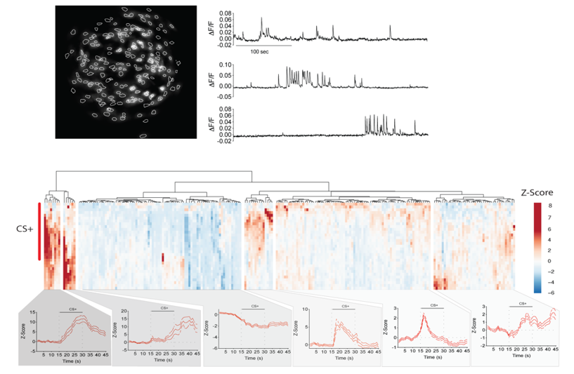
In Vivo Calcium Imaging in Freely Moving Mice
Analysis of single-cell calcium activity in freely moving laboratory animals has advanced our ability to understand how distinct neuronal ensembles represent cognitive, sensory and affective states. Such approaches have led to deep understanding of how neurons encode distinct behavioral states and how pharmacological interventions affect this process to exert therapeutic actions. In vivo brain imaging approaches have the potential to broadly increase our understanding of the neural mechanisms underlying essential biobehavioral processes of psychiatric disorders.
Feinberg Research Resources
The Feinberg Office of Research works with units across Northwestern to support investigators' research at every step of the process.