
February 2026 Newsletter
February 2026 Newsletter
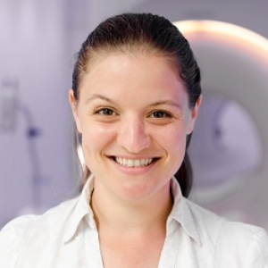
Faculty Profile
Molly Bright, DPhil, is an assistant professor of Physical Therapy and Human Movement Sciences and of Biomedical Engineering in the McCormick School of Engineering. Her research focuses on developing functional MRI (fMRI) techniques to assess the function and interaction of neural activity and vascular physiology throughout the central nervous system, with an emphasis on translating these approaches directly to patients with movement impairments.
What are your research interests?
I develop fMRI methods to understand the human brain, with a special focus on people with movement impairments. I am particularly interested in capturing neural activity throughout the entire central nervous system – lots of studies only capture cortical information, but this is only one part of the larger story when thinking about human movement. My lab works hard to get high-quality data in the brainstem, cerebellum and even spinal cord to really achieve a “systems” view of motor control. I also want to push these methods to get trustworthy information in individual patients, rather than relying on averaging our data across larger populations. Our research studies focus on “precision” imaging, changing how we acquire and process fMRI data to map brain function of a single subject. Finally, fMRI is fundamentally a vascular imaging method, where we can infer neural activity. I try to capitalize on this “dual nature” and characterize both neural and vascular function. This is particularly important in pathologies where both systems are impacted, like in stroke.
What is the ultimate goal of your research?
I want fMRI to be useful. It is a technique with a lot of potential and a lot of use in research studies, but it has yet to fully translate and make meaningful clinical impact. I want to help get us over that hurdle and make fMRI a reliable tool that can contribute to clinical decision-making. For example, I want fMRI to guide the optimization and even personalization of rehabilitative treatments.
How did you become interested in this area of research?
With my background in physics, I became interested in medical imaging quite early on, and I entered a unique graduate training program that linked the University of Oxford and the National Institutes of Health. Speaking with potential mentors there, I thought MRI was a particularly exciting opportunity because this one scanner could be sensitive to an incredibly diverse range of physiological factors. With one tool, you could see overt structural brain changes, subtle tissue damage, impaired perfusion, metabolic changes, iron deposition and even localized neural activity. It’s also endlessly fascinating to educate myself about neuroscience and physiology while I adapt MRI methods to study the brain.
More practically, it is a research field where I enjoy getting to wear many hats, with any given day involving some coding and simulations, hardware design and testing, signal processing, physiological modeling, clinical trial oversight, working with colleagues from all over the medical school, and engaging with our research participants. It’s genuinely fun.
What types of collaborations are you engaged in across campus (and beyond)?
My most productive collaborations here in Chicago are with physical therapy investigators at Northwestern and the Shirley Ryan AbilityLab, combining my imaging methodology with their expertise in movement impairments and rehabilitative interventions. With Julius Dewald, PhD, we are mapping the neural control of movement in chronic stroke, seeking to characterize the specific motor pathways that a given individual is using. With Milap Sandhu, PhD, we are using neuroimaging to identify the mechanisms by which acute intermittent hypoxia therapy improves strength and walking function in multiple sclerosis, with the goal of using imaging to one day personalize this intervention.
Where have you recently published papers?
Much of our methods development work finds a home in Imaging Neuroscience and similar imaging-focused journals. Other recent studies have been published in Cerebral Cortex, the Journal of Cerebral Blood Flow and Metabolism, Scientific Reports and IEEE publications.
Who inspires you?
I’m currently teaching a graduate course in physiology for biomedical engineers, and I’ve been incorporating historic literature into the reading assignments. These papers, many from the early 1900s, reflect an entirely different landscape of research, where scientists had to be incredibly creative to achieve their experiments without modern technology. The “giants” of that time spawned multiple new research disciplines, leading to countless medical advances. I’ve found myself quite inspired by these historical scientists, and I want to emulate their openness and curiosity about how the world works.
