Presenting Author:
Principal Investigator:
Wendy Murray, Ph.D.
Department:
Physical Medicine and Rehabilitation
Keywords:
Skeletal Muscle, Ultrasonography, Fascicle length, Upper Limb
Location:
Third Floor, Feinberg Pavilion, Northwestern Memorial Hospital
B157 - Basic Science
Extended Field-of-View Ultrasound in the Extensor Carpi Ulnaris
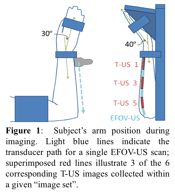
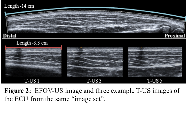
INTRODUCTION Fascicle length is an important parameter in defining a muscle’s force generating capacity. Currently, ultrasound is the most common method of measuring fascicle length in vivo [1]. However, 24 of 26 forearm muscles have fascicles longer than the field-of-view of common traditional ultrasound (T-US) probes. As such, little work has been done to quantify in vivo forearm muscle architecture. The extended field-of-view ultrasound (EFOV-US) algorithm fits together a sequence of T-US images obtained via a continuous ultrasound scan [2], facilitating direct measurement of longer, curved fascicles. The objective of this study was to determine the validity and reliability of the EFOV-US method for obtaining fascicle lengths in the extensor carpi ulnaris (ECU). METHODS To evaluate the validity of the measurement protocol used in this study, an ultrasound phantom was imaged to compare measurements of objects made using a caliper and those made using EFOV-US images of the phantom. To determine in vivo validity and reliability of EFOV-US’s long scans, the right arms of 8 healthy subjects (4F/4M, ages 23-29) were imaged using both EFOV-US and T-US. The subject’s arm was placed in a joint posture that shortened the ECU to the extent that entire fascicle lengths could be captured within a single T-US image (Fig. 1). The sonographer obtained an EFOV-US image of the ECU followed by 6 T-US images that spanned the length of the ECU (Fig. 2). RESULTS The measurements from the ultrasound phantom indicate that the EFOV-US imaging and analysis methods produced valid results. We observed an average error of 2.2 ± 1.3mm (4.6% average phantom object length). A Bland-Altman plot demonstrated agreement between direct caliper and EFOV-US measurements of the phantom objects (bias= -0.67mm, 95% CI: -5.57 to 4.23mm). EFOV-US fascicle measurements are comparable to those obtained from T-US, a well-established method. Average EFOV-US fascicle lengths (27.9 ± 3.7mm) were not significantly different (p=0.18) than T-US fascicle lengths (28.2 ± 4.0mm). Bland-Altman analysis also indicated that the two methods agree (bias= -0.35mm, 95% CI:-1.47 to 0.77mm). Between trial reliability (ICC=0.99 95% CI: 0.95-1.00) was excellent [3]. CONCLUSION Only one previous study has quantified in vivo fascicle lengths in a forearm muscle, despite the ubiquity of ultrasound studies in other muscle groups and the importance of architecture in understanding muscle force generation. The EFOV-US method has the capacity to capture the entire length of the fascicle in one image; this is advantageous in the forearm where over 90% of muscles have optimal fiber lengths longer than the field-of-view of T-US. Utilizing this method in patient populations may lead to improvements in diagnosis, surgery guidance, and rehabilitation techniques. REFERENCES 1. Kwah,L. et al.J App Physiol 114,761-69,2013. 2. Weng,L. et al.Radiology 203,877-880,1997. 3. Noorkoiv,M. et al.J App Physiol 3,627-34,2010.