Presenting Author:
Elias Gounaris, Ph.D.
Principal Investigator:
David Bentrem, M.D.
Department:
Surgery
Keywords:
colon cancer, cathepsin, NIRF endoscope, NIRF probes, in vivo fluorescence imaging
Location:
Third Floor, Feinberg Pavilion, Northwestern Memorial Hospital
B173 - Basic Science Women's Health Research
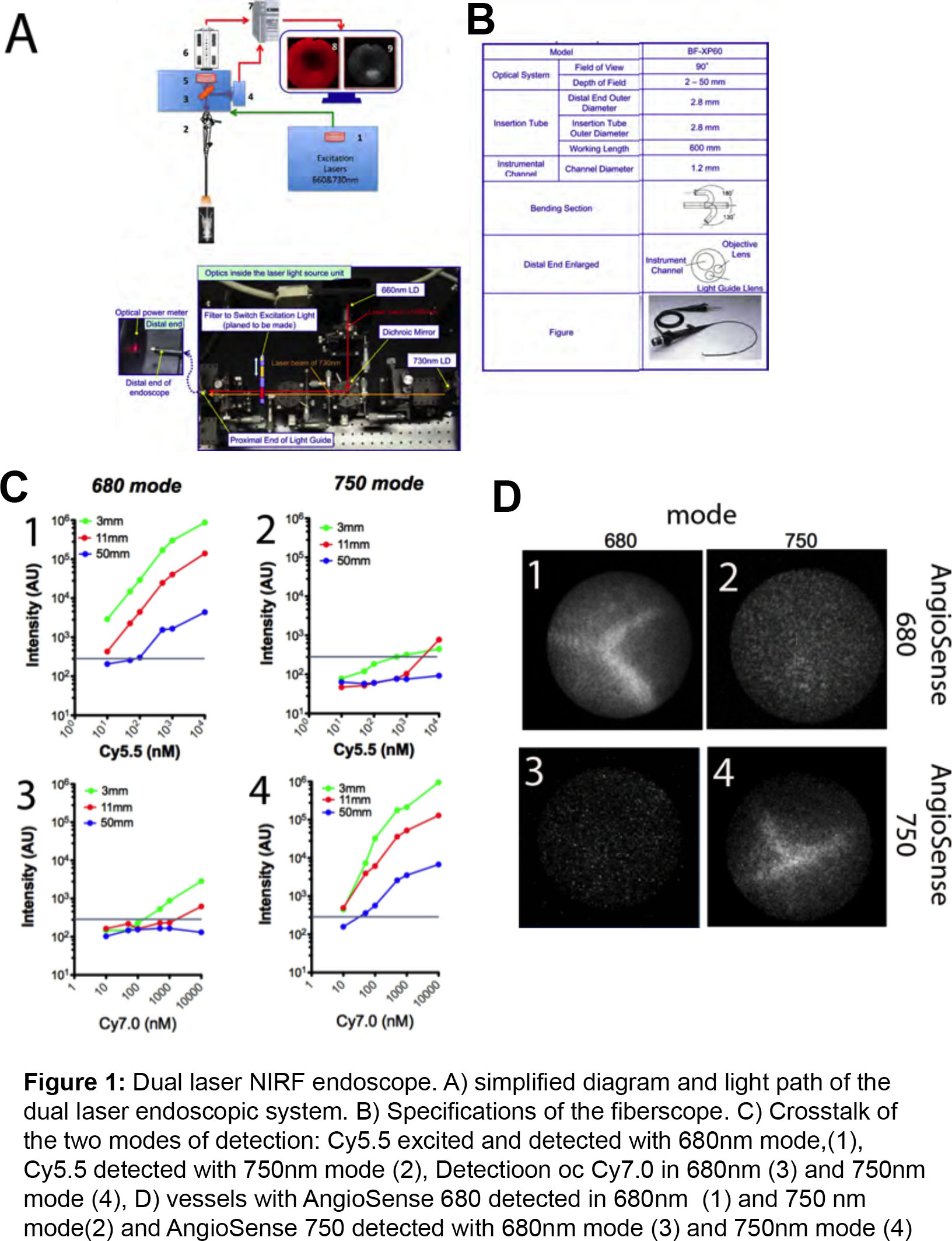
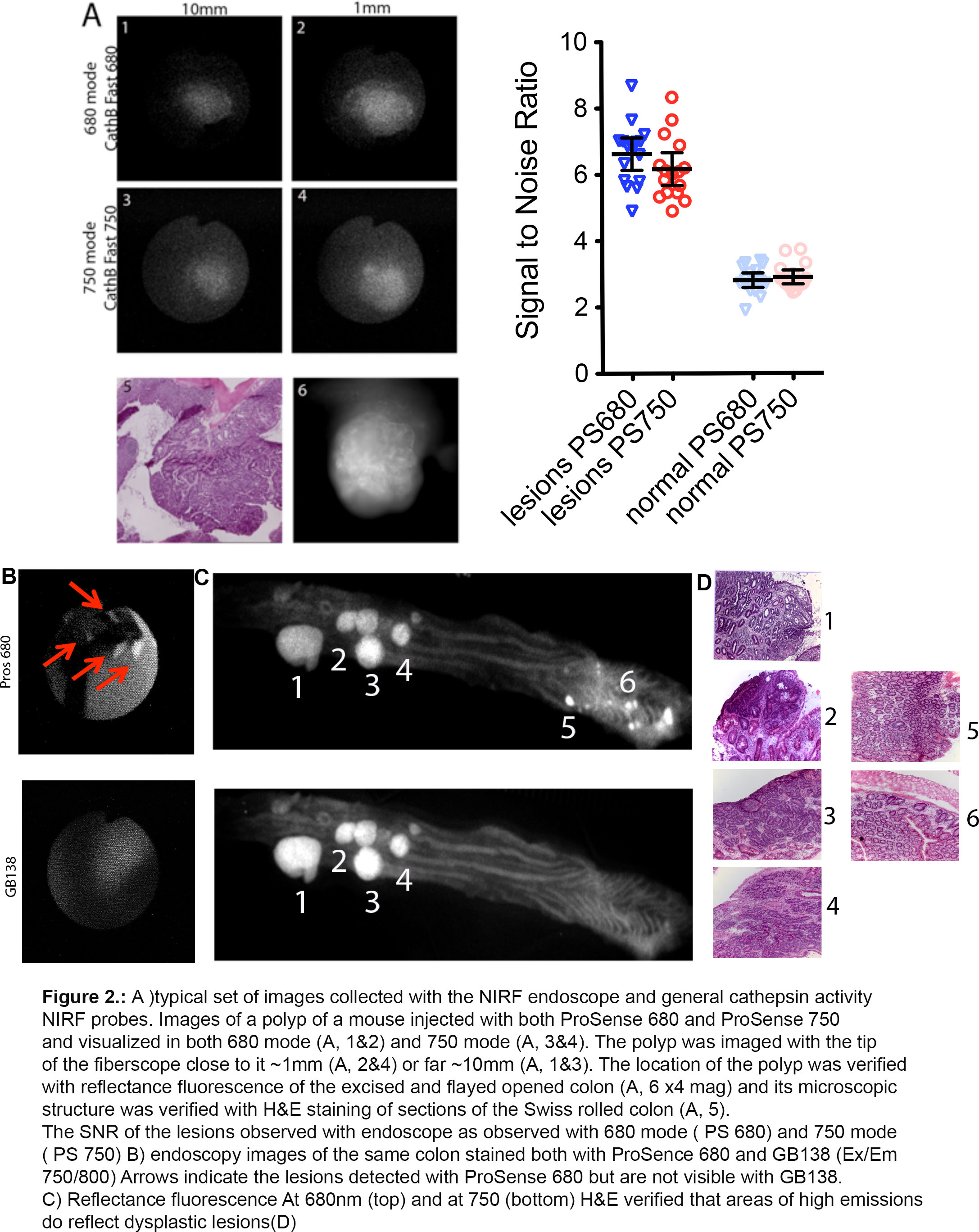
Dual Laser NIRF Endoscope to Compare NIRF Probes in vivo
A dual laser near infrared fluorescence endoscope was developed in collaboration with Olympus (Tokyo, Japan). The intended purpose of this system was to permit in vivo comparison studies using multiple NIRF cathepsin probes for simultaneous in vivo interrogation of multiple biological processes. This system was intended to a) permit two independent examination modes (namely at 680nm and 750nm wavelengths), b) have minimal cross talk between the two examination modes, c) produce well-adjusted excitation light energy such that the two examination modes would be evenly weighted, d) operate with a number of fiberscopes with diverse specifications and finally e) permit image recording at video frequency (10 frames/second). Our system can image autochthonous grown tumors in the gastrointestinal tract, utilizing two modes of detection. There is very limited cross talk between the two modes. The cross talk is only possible when the tip of the fiberscope is very close to the lesion and the concentration of the NIRF probes is very high. The two modes are balanced (excitation power is similar). One of the most important features of the system is that it is possible to utilize various fiberscopes that can access other tissues of the mice like esophagus and lung in non-invasive mode, as well as, pancreas, prostate and breast tissues laparoscopically. This gives us the possibility to use specific NIRF probes to detect specific disorders. We developed the dual NIRF laser light source for the new NIRF endoscope to address the selection of the suitable NIRF cathepsin probes to detect early dysplastic lesions11. There is a report, which is trying to address the problem of comparing the ABPs with SBPs. They have used nude mice, which subcutaneously injected with tumor cells. In this report, ABPs look more efficient to detect the tumors. Injected tumors, as compared to the autochthonous tumors, posing problems as they a) growing very fast without adequate vascular system b) the center of the tumor is necrotic, c) inflammatory myeloid cells are located only in the periphery of the tumors13. Our NIRF endoscopic system is designed to overcome all these problems, as it uses mice that grow tumors automatically, in the tissue localization, showing a marked increase of the vascularity, and the inflammatory myeloid cells are integrated into the tumor’s stroma. In an effort to permit rigorous co-registered comparison studies and also the possibility for salient combinations of different NIRF probes during a single examination, we recently developed a new dual laser multi-channel NIRF endoscope. Herein we report the development and systemic evaluation of this new NIRF endoscope along with initial studies using this dual laser endoscope for comparisons of various probes in the same lesions in transgenic mouse model of colon cancer