Presenting Author:
Danijela Maric, Ph.D.
Principal Investigator:
Thomas Hope, Ph.D.
Department:
Cell and Molecular Biology
Keywords:
HIV/SIV, Rhesus Macaque, Rectal Mucosa, Th17
Location:
Third Floor, Feinberg Pavilion, Northwestern Memorial Hospital
PH1 - Public Health & Social Sciences Women's Health Research
Defining HIV/SIV Infection After Rectal Challenge in Rhesus Macaque Model
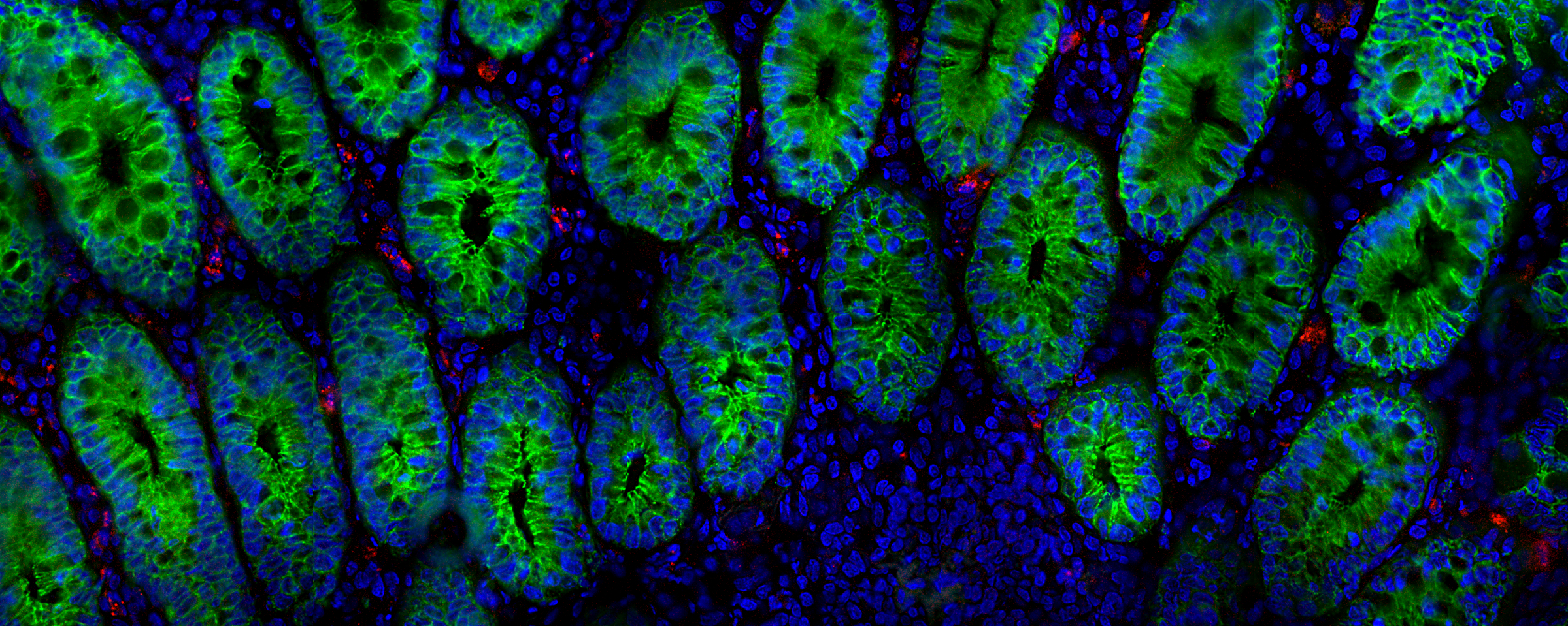
Receptive anal intercourse accounts for more than half of new HIV-1 infections within the United States each year; yet, rectal transmission of HIV is an understudied area. Developing a working knowledge of the initial barriers and cellular targets of HIV could lead to the development of more effective strategies to prevent rectal transmission. Four rhesus macaques (RMs) were rectally challenged with a single-round dual-reporter SIV virus that expresses Luciferase and near-infrared fluorescent protein 670 (iRFP670) that was pseudotyped with an envelope protein from a CCR5 tropic HIV virus, JR-FL. Forty-eight hours after the challenge anal and rectal tissue were removed and analyzed with In Vivo Imaging System to detect the foci of infection. Tissue exhibiting robust luminescent signal were cryosectioned to confirm infection by fluorescence microscopy, spectral imaging and nested PCR techniques. Subsequently infected cells were phenotyped by chemokine receptor staining and RNA-fluorescent in situ hybridization (RNA-FISH) to monitor expression of mRNA transcripts. We reliably detected infected cells 48-hours post challenge throughout the anal and rectal tissues. To phenotype the infected cells we stained for CD4 and found that all iRFP670 and Luciferase expressing cells were CD4 positive. We next examined expression of CD3 and found out that 68% of infected cells were CD3 positive, consistent with our previous study. This indicates that significant portions (32%) of CD3 negative cells, including macrophages and dendritic cells (DCs) are transduced shortly after exposure. To determine if CCR6+ T-cells are early targets of SIV, we co-stained tissue sections with CD3 and CCR6 antibodies, as CD3+/CCR6+ T-cells were recently identified to be preferentially targeted by SIV after vaginal challenge. Our to date analysis indicates that 54% of transduced cells are CD3+/CCR6+ (Th17 T-cells), 39% are CD3-/CCR6+ cells (immature dendritic cells) and 7% are CD3-/CCR6- cells (macrophages). Our current experimental efforts are focused on rectal co-challenges with our Luciferase reporter virus and WT SIV with a goal of using our reporter virus as a tool to find cells infected by the real virus. Together these studies will allow us to determine the cellular targets of SIV/HIV and will lay the groundwork for our future work, in which we hope to examine the efficacy of various antiretroviral drugs in preventing transmission at this portal of entry.