Presenting Author:
Janushi Dalal, M.D.
Principal Investigator:
Janushi Dalal, M.D.
Department:
Radiology
Keywords:
Breast Imaging, Dermal Lesions, Malignant Potential
Location:
Ryan Family Atrium, Robert H. Lurie Medical Research Center
C126 - Clinical Women's Health Research
Evaluation of dermal lesions of the breast
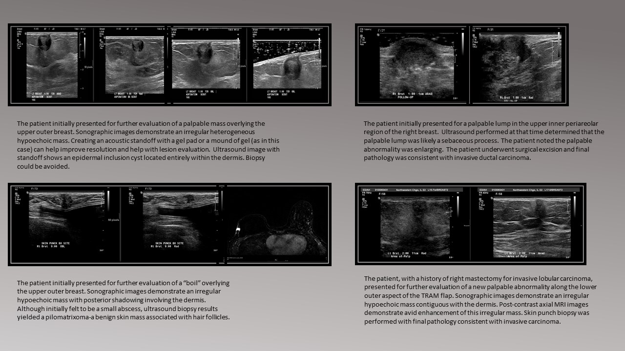
Breast imagers often are required evaluate dermal lesions. Lesions which arise in the dermal layer are often considered benign, with the vast majority representing epidermal inclusion cysts, sebaceous cysts, and vascular tumors. Typically these lesions are managed clinically and undergo routine surveillance. However, malignant and pre-malignant entities can also occur in this superficial location. Therefore, it is essential for a breast imager to be familiar with the underlying dermal anatomy as management of such lesions is often dependent upon the location. Lesions which arise in the hypodermis (subcutaneous fat) are usually benign. It is important to remember that superficial breast cancers can also arise in the hypodermal layer as terminal duct lobular units can occasionally be present. It is crucial that these masses are not classified as benign simply because of the anterior location. It is critical to use the BIRADS sonographic imaging features to characterize these masses to further determine management. The anatomy of the areola is unique. Lactiferous ducts are present associated with sebaceous glands. These glands may open directly on to the areola. Legions within the nipple-areola complex are not considered to be extra parenchymal because of this direct extension to the skin. The glands, which are located just deep to the areolar muscle may develop any breast abnormalities including malignant processes. Learning Objectives 1. Understand the basic anatomy of the breast dermis 2. Identify the common and rare lesions which may occur in each dermal layer 3. Explain complex and varying anatomy of the nipple areolar complex 4. Demonstrate the utility of the BIRADS for determining evaluation and management of dermal lesions. Abstract Content: In this presentation, we will identify both common and atypical sonographic findings associated with breast dermal lesions using individual cases. This will include both a schematic review as well as actual cases reviewed at our institution. In total, ten cases will be reviewed including work-up methodology and final assessment Corresponding pathologic images will also be included. Conclusion: Although dermal lesions of the breast are typically benign, understanding the complex anatomy is crucial in order to identify and manage early breast carcinomas.