Presenting Author:
Principal Investigator:
Kalliopi Siziopikou, M.D.
Department:
Pathology
Keywords:
Pregnancy-associated breast cancer, PD-L1, PD-1, tumor infiltrating lymphocytes
Location:
Ryan Family Atrium, Robert H. Lurie Medical Research Center
C81 - Clinical Women's Health Research
PD-L1 Expression in Tumor Infiltrating Lymphocytes in Pregnancy-Associated Breast Cancer
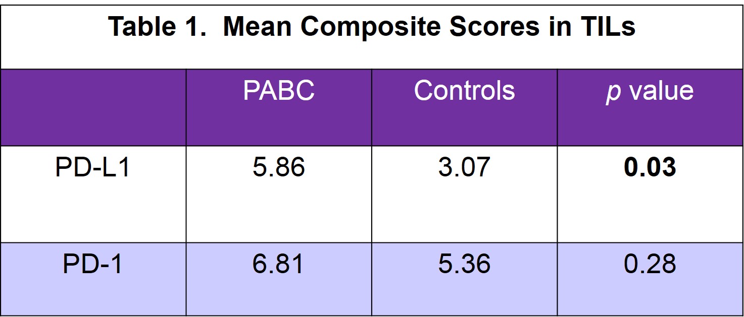
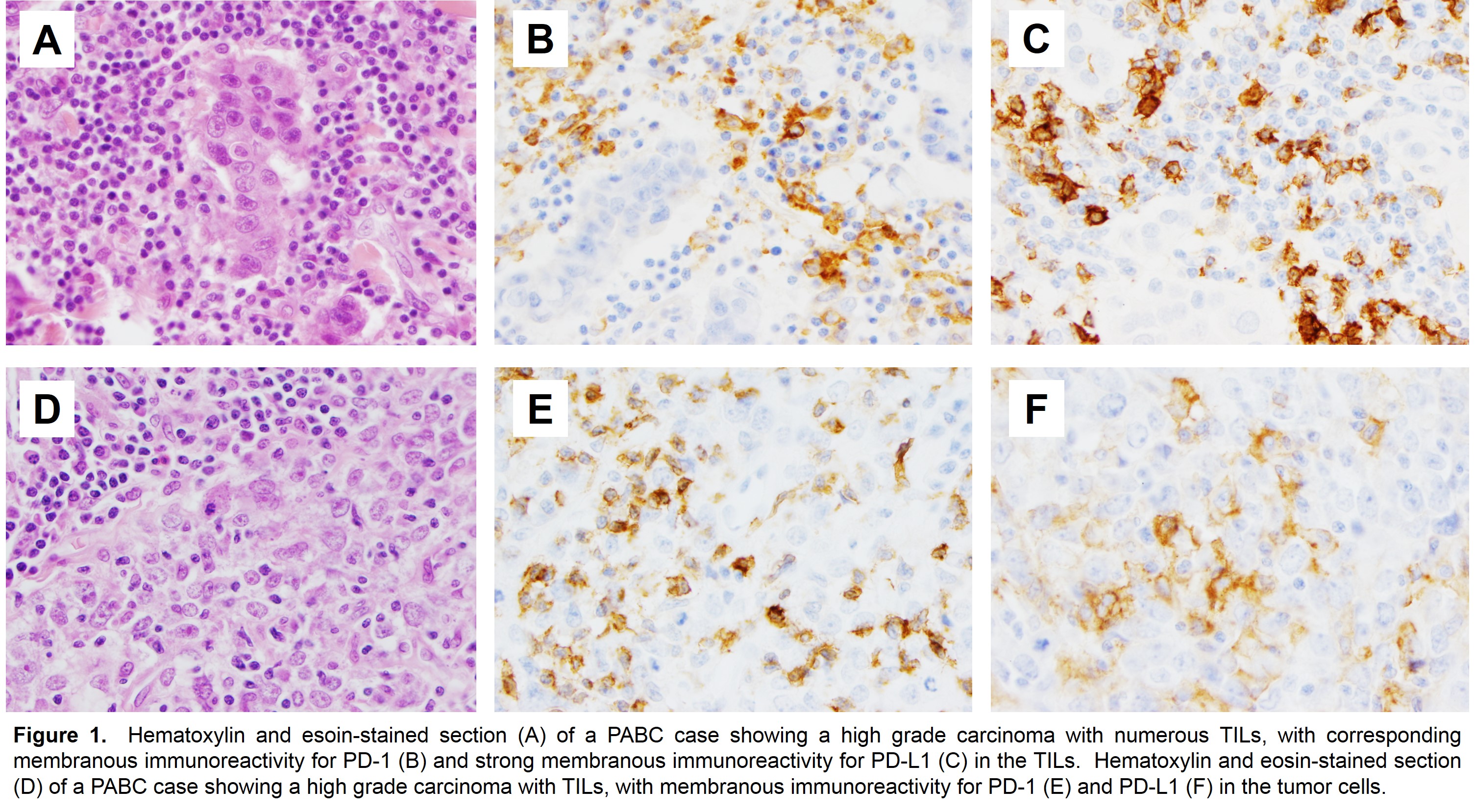
Summary: Pregnancy associated breast cancer (PABC), diagnosed during or after gestation, is triple negative and associated with a poor prognosis. We previously assessed the immune microenvironment of invasive breast carcinomas in young women and reported that tumor infiltrating lymphocytes (TILs) were more prominent in PABC. Programmed cell death protein 1 (PD-1) is upregulated following activation of lymphocytes, while programmed death ligand 1 (PD-L1) is one of the primary ligands that it interacts with to inhibit T-cell activation and proliferation. PD-L1 may also be constitutively expressed on tumor cells as a result of oncogenic signaling or epithelial-mesenchymal transition. Emerging evidence suggests that the local immune system, particularly the interactions between PD-1 and PD-L1, is also key in breast cancer progression and in breast tumor responses to chemotherapy and targeted therapy. Objective: In this study, our goal was to assess the expression of PD-1 and PD-L1 in both TILs and tumor cells in PABC and in age-/stage-/grade-matched nulliparous women and to correlate their expression with clinicopathologic characteristics in this aggressive type of breast carcinomas. Methods: 21 patients diagnosed with PABC within two years of pregnancy (mean age=35.7, range=26-48) and 15 matched controls (mean age=37.5, range=29-51) were evaluated. Immunohistochemical stains for PD-1 and PD-L1 were performed. Extent (1=1-25% positive tumor cells, 2=26-50%, 3=51-75%, 4=76-100%) and intensity (1=weak, 2=moderate or 3=strong) were assessed and a composite score (CS) calculated by multiplying the extent by intensity (range=0-12; weak=1-3; moderate=4-8 and strong=9-12). Results: The mean CS for PD-L1 in TILs was significantly higher in PABC (5.86) compared to controls (3.07), p=0.03. Further, strong expression of PD-L1 in TILs was only observed in PABC (9/21, 42.9%); none of the controls had strong PD-L1, p=0.01. The high expression of PD-L1 in PABC TILs was independent of tumor grade, hormone receptor and HER2 status, and other histologic features including lymph node metastasis. Expression of PD-1 in TILs was similar in both PABC and controls (mean CS 6.81 and 5.36, respectively). Immunoreactivity in the tumor cells themselves was rare with only two PABC and four control cases expressing PD-1 and PD-L1. Conclusions: 1. TILs in PABC have significantly higher PD-L1 expression. 2. Strong expression of PD-L1 in TILs was only observed in PABC. 3. High PD-L1 expression in TILs was independent of the tumor characteristics in this series. 4. PD-1 is expressed similarly in TILs in both PABC and controls. 5. Rare cases may have PD-1 and PD-L1 expression in the tumor cells themselves. The results showing significant expression of PD-L1 in PABC TILs add to the understanding of the role of the microenvironment in breast cancer progression. These complex interactions between tumors cells and the local immune system may predict response to targeted therapy.